d o t o r e g . c o m
Dr. Mete ALPASLAN
VPC-like Artifacts

ECG 1. Above is a 6-channel ECG from a 35 years-old apparently healthy man with no known heart disase.
Short-lasting baseline drift gives the appearence of wide QRS and hence ventricular premature contraction (VPC) at first glance.
However these beats are not premature.
The short-lasting baseline drift also has not affected the T wave vector (which is expected in a VPC).
Additionally, lead I is devoid of baseline drift and the narrow QRS has a normal T wave.
These clues show that they are not VPCs.
Click here for a more detailed ECG

ECG 2. The ECG above was recorded during treadmill exercise test (stress test).
Short-lasting baseline drift gives the appearence of wide QRS and hence ventricular premature contraction (VPC) at first glance.
However, these are not VPCs, but baseline drift giving the impression of a VPC.
The adjacent narrow QRS complexes also show that the normal QRS complexes are present at the usual expected site.
Click here for a more detailed ECG

ECG 3. The ECG above, is from a 22 years-old healthy man with a normal ECHOcardiogram.
Limb leads show VPC-like artifact.
Click here for a more detailed ECG

ECG 4. The ECG above shows a VPC-like artifact.
Click here for a more detailed ECG

ECG 5. In the above 2-channel Holter recording, baseline drift artifact is falsely perceived as R-on-T
by the ECG machine's computer. The artifact perceived as ventricular beats are marked as V.
In fact, there are no VPCs, no R-onT.
Click here for a more detailed ECG

ECG 6. Short-lasting baseline shift mimicks QRS widening and hence VPC.
In fact, there is no premature beat.
RR intervals are similar. This is a VPC-like artifact.
Click here for a more detailed ECG

ECG 7. Short-lasting baseline shift mimicks QRS widening and hence VPC.
In fact, there is no premature beat.
RR intervals are similar. This is a VPC-like artifact.
Click here for a more detailed ECG

ECG 8. The rhythm of the above ECG is atrial fibrillation.
Short-lasting baseline shift mimicks QRS widening and hence VPC.
In fact, there is no premature beat.
Click here for a more detailed ECG

ECG 9. The rhythm of the above ECG is sinus.
Short-lasting baseline shift mimicks QRS widening and hence VPC.
In fact, there is no premature beat.
Click here for a more detailed ECG

ECG 10. Above is an ECG from a hypertensive patient with renal failure. The rhythm is sinus tachycardia.
Short-lasting baseline shift mimicks QRS widening and hence VPC.
In fact, there is no premature beat.
Click here for a more detailed ECG
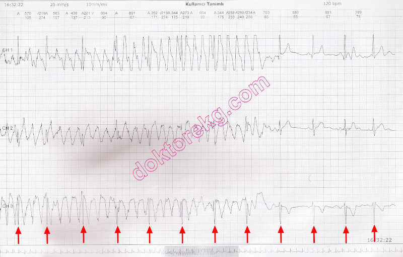
ECG 11. Above is a 3-channel tracing from an ambulatory ECG (rhythm Holter) monitoring. The rhythm may seem like ventricular tachycardia, at first glance. The patient's response to itching induced by the electrode may cause artifact mimicking VPCs (or VT as in the above tracing). Careful inspection of the tracing reveals almost regular QRS complexes.
Click here for a more detailed ECG
ECG 1. Above is a 6-channel ECG from a 35 years-old apparently healthy man with no known heart disase.
Short-lasting baseline drift gives the appearence of wide QRS and hence ventricular premature contraction (VPC) at first glance.
However these beats are not premature.
The short-lasting baseline drift also has not affected the T wave vector (which is expected in a VPC).
Additionally, lead I is devoid of baseline drift and the narrow QRS has a normal T wave.
These clues show that they are not VPCs.
Click here for a more detailed ECG
ECG 2. The ECG above was recorded during treadmill exercise test (stress test).
Short-lasting baseline drift gives the appearence of wide QRS and hence ventricular premature contraction (VPC) at first glance.
However, these are not VPCs, but baseline drift giving the impression of a VPC.
The adjacent narrow QRS complexes also show that the normal QRS complexes are present at the usual expected site.
Click here for a more detailed ECG
ECG 3. The ECG above, is from a 22 years-old healthy man with a normal ECHOcardiogram.
Limb leads show VPC-like artifact.
Click here for a more detailed ECG
ECG 4. The ECG above shows a VPC-like artifact.
Click here for a more detailed ECG
ECG 5. In the above 2-channel Holter recording, baseline drift artifact is falsely perceived as R-on-T
by the ECG machine's computer. The artifact perceived as ventricular beats are marked as V.
In fact, there are no VPCs, no R-onT.
Click here for a more detailed ECG
ECG 6. Short-lasting baseline shift mimicks QRS widening and hence VPC.
In fact, there is no premature beat.
RR intervals are similar. This is a VPC-like artifact.
Click here for a more detailed ECG
ECG 7. Short-lasting baseline shift mimicks QRS widening and hence VPC.
In fact, there is no premature beat.
RR intervals are similar. This is a VPC-like artifact.
Click here for a more detailed ECG
ECG 8. The rhythm of the above ECG is atrial fibrillation.
Short-lasting baseline shift mimicks QRS widening and hence VPC.
In fact, there is no premature beat.
Click here for a more detailed ECG
ECG 9. The rhythm of the above ECG is sinus.
Short-lasting baseline shift mimicks QRS widening and hence VPC.
In fact, there is no premature beat.
Click here for a more detailed ECG
ECG 10. Above is an ECG from a hypertensive patient with renal failure. The rhythm is sinus tachycardia.
Short-lasting baseline shift mimicks QRS widening and hence VPC.
In fact, there is no premature beat.
Click here for a more detailed ECG
ECG 11. Above is a 3-channel tracing from an ambulatory ECG (rhythm Holter) monitoring. The rhythm may seem like ventricular tachycardia, at first glance. The patient's response to itching induced by the electrode may cause artifact mimicking VPCs (or VT as in the above tracing). Careful inspection of the tracing reveals almost regular QRS complexes.
Click here for a more detailed ECG