d o t o r e g . c o m
Dr. Mete ALPASLAN
Sinus Arrest - Sinus Pause - Sinus Standstill
Diagnostic criteria
 Sinus node cannot produce an impulse at the expected time. On the ECG, this is seen as a long isoelectric line between the last QRS complex and the first P wave following the pause.
In sinus arrest (sinus standstill), the pause is NOT a multiple
of the P-P interval that preceded the pause. In sinoatrial exit block, the pause is a multiple of the P-P interval that preceded the pause.
If sinus arrest lasts long enough, then atrial, nodal or ventricular escape beat (or rhythm) may be seen. If no escape beat or rhythm ensues, then it becomes cardiac arrest.
Remember: A blocked atrial premature beat may mimick sinus arrest, at first glance. In this case, we expect to see a blocked P wave deforming its preceding T wave.
Causes
Increased vagal tone
Carotid sinus hypersensitivity
Sleep apnea
Sick sinus syndrome
Myocardial ischemia / infarction
References (with links to similar ECGs)
Can J Rural Med 2012;17:110-113 - Answer.
Int Arch Med 2012;5:11.
Korean J Anesthesiol 2010;59(Suppl):S137-S140.
BMJ Case Rep. 2011 Apr 13;2011. pii: bcr1120103519.

ECG 1. Sinus arrest in an adult. After the first 5 P waves no P waves follow at the expected time . The first P wave after the
sinus arrest came lately.
Click here for a more detailed ECG

ECG 2. Sinus arrest in a 3 years old boy. Children have a higher resting heart rate when compared to adults. After the first 3
P waves there comes P wave at the expected time . The PP interval of the first P wave after the sinus arrest is NOT a
multiple of the P-P interval that preceded the pause (there is no sinoatrial exit block) Sinus arrest can be due to increased
vagal tone.
Pediatric cardiologist Dr. Mahmut Gokdemir has donated this ECG to our website.
Click here for a more detailed ECG

ECG 3a. The tracing above has been recorded 30 seconds before sinus arrest during a passive tilt table test.
Heart rate is 56/minute.
This ECG has been donated by Dorota Zysko.
Click here for a more detailed ECG

ECG 3b. The same patient's tracing 15 seconds before sinus arrest shows sinus bradycardia. Heart rate is 37/minute.
This ECG has been donated by Dorota Zysko.
Click here for a more detailed ECG

ECG 3c. The same patient during vasovagal syncope at the 24th minute of the passive tilt table test. The rhythm tracing shows
sinus arrest longer than 5 seconds. The resultant decrease in cerebral perfusion results in jerking movements which in turn
caused artifacts .
This ECG has been donated by Dorota Zysko.
Click here for a more detailed ECG

ECG 3d. The heart rate increased to 44/minute 15 seconds after syncope.
This ECG has been donated by Dorota Zysko.
Click here for a more detailed ECG

ECG 4a. The ECG above belongs to a 74 years-old woman complaining of dizziness. The ECG is normal.
She has normal coronary arteries.
Click here for a more detailed ECG

ECG 4b. The same patient's ECG (limb leads) on a different day shows sinus arrest .
Click here for a more detailed ECG

ECG 4c. The same patient's ECG (chest leads) on a different day shows sinus arrest .
Click here for a more detailed ECG

ECG 4d. The above tracings are from the Holter recording of the same patient. This time, there are longer periods of sinus arrest.
In both tracings above, the first 4 P waves are regular . Then a sinus arrest of 2.5 seconds occur in the upper tracing and
a nodal escape beat arises which has no P wave preceding it . In the bottom tracing the pause is longer this time (3.7 seconds).
Again the sinus arrest is long enough and a nodal escape beat arises which has no P wave preceding it .
Click here for a more detailed ECG

ECG 5a. Above is an ECG from a 51 years-old woman. The ECG shows no clear abnormality.
She was admitted to Cardiology clinic with the complaints of attacks of palpitation and near-syncope.
For the last 2 years, she was taking Metoprolol tablets.
A normal ECHOcardiogram ruled out heart failure and valvular heart disease.
Metoprolol was stopped and Holter (ambulatory cardiac rhythm) monitor was requested.
Click here for a more detailed ECG

ECG 5b. The above rhythm tracing is from her Holter recording (while not taking Metoprolol).
A pause of 2.4 seconds of duration was recorded at 07:36 AM (while she was awake).
Click here for a more detailed ECG

ECG 5c. The above rhythm strips are also from her Holter recording.
Sudden increases in heart rate are seen: Atrial tachycardia following sinus braycardia.
Click here for a more detailed ECG

ECG 5d. The two Holter strips above belong to the same patient..
The strips show sinus pauses, each lasting more than 5 seconds. Both pauses follow atrial tachycardias.
Click here for a more detailed ECG

ECG 5e. Above Holter rhythm tracings also belong to the same patient.
Sudden increases and decreases of heart rate are seen.
Consecutive bradycardias and atrial tachycardias: bradycardia-tachycardia syndrome.
Click here for a more detailed ECG
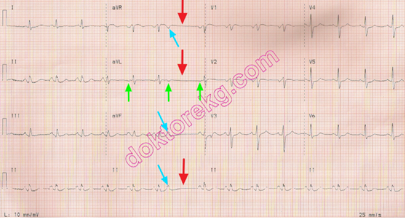
ECG 6. The above ECG is from a 58 years-old woman with chronic hypertension.
It may look like sinus arrest, at first glance.
However, the pause is due to a blocked atrial premature beat.
The blocked atrial premature beat deforms the preceding T wave.
The simultaneously recorded lead aVR (where T waves are isoelectric in this patient) shows atrial depolarizaton waves clearly.
Click here for a more detailed ECG

ECG 7a. Above is an ECG from a middle-aged woman.
She was transferred to our hospital with the diagnosis of sinus arrest.
What is your diagnosis? Is there a real pause?
Dr. Sinan Altan Kocaman has donated the above ECG to our website.
Click here for a more detailed ECG
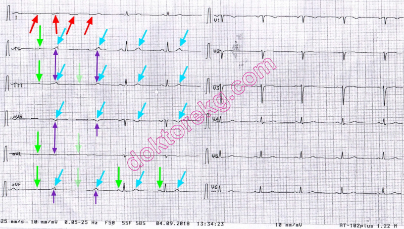
ECG 7b. Above is the explanation for the same ECG:
The deflections mimicking P waves on the left side are in fact T waves.
If they are T waves, then where are their preceding QRS complexes? They are not seen.
It is "not because that they do not exist". They are not depicted because of the artifact.
There are 3 clues to suggest artifact in this patient:
1- T waves cannot be formed without preceding QRS complexes.
2- Lead I shows baseline drift due to artifact.
3- Also, this artifact deforms the simultaneous P waves resulting in the appearence of subtle deflections.
The second P wave from the left side is not seen where it should be.
In conclusion, there is no sinus pause in this ECG, but artifact mimicking pause.
Her heart rate is 58/minute.
Dr. Sinan Altan Kocaman has donated the above ECG to our website.
Click here for a more detailed ECG

ECG 8. The above ECG belongs to a 13 years-old boy with frequent atrial premature beats.
Atrial premature beats deform the preceding T waves.
Some atrial premature beats are conducted with aberration resulting in wide QRS complexes.
These wide QRS complexes are not ventricular premature contractions.
There are two reasons for this: T waves preceding the wide QRS complexes are different
(due to the deforming effect of the atrial premature beat); also there is no compensatory pause.
The seemingly sinus arrest is in fact due to a blocked atrial premature beat.
There is no sinus arrest.
Arm electrodes were inadvertently reversed in this ECG
Prof. Dr. Nazlıhan Günal has donated the above ECG to our website.
Click here for a more detailed ECG
Diagnostic criteria
Sinus node cannot produce an impulse at the expected time. On the ECG, this is seen as a long isoelectric line between the last QRS complex and the first P wave following the pause.
In sinus arrest (sinus standstill), the pause is NOT a multiple
of the P-P interval that preceded the pause. In sinoatrial exit block, the pause is a multiple of the P-P interval that preceded the pause.
If sinus arrest lasts long enough, then atrial, nodal or ventricular escape beat (or rhythm) may be seen. If no escape beat or rhythm ensues, then it becomes cardiac arrest.
Remember: A blocked atrial premature beat may mimick sinus arrest, at first glance. In this case, we expect to see a blocked P wave deforming its preceding T wave.
Causes
Increased vagal tone
Carotid sinus hypersensitivity
Sleep apnea
Sick sinus syndrome
Myocardial ischemia / infarction
References (with links to similar ECGs)
Can J Rural Med 2012;17:110-113 - Answer.
Int Arch Med 2012;5:11.
Korean J Anesthesiol 2010;59(Suppl):S137-S140.
BMJ Case Rep. 2011 Apr 13;2011. pii: bcr1120103519.
ECG 1. Sinus arrest in an adult. After the first 5 P waves no P waves follow at the expected time . The first P wave after the
sinus arrest came lately.
Click here for a more detailed ECG
ECG 2. Sinus arrest in a 3 years old boy. Children have a higher resting heart rate when compared to adults. After the first 3
P waves there comes P wave at the expected time . The PP interval of the first P wave after the sinus arrest is NOT a
multiple of the P-P interval that preceded the pause (there is no sinoatrial exit block) Sinus arrest can be due to increased
vagal tone.
Pediatric cardiologist Dr. Mahmut Gokdemir has donated this ECG to our website.
Click here for a more detailed ECG
ECG 3a. The tracing above has been recorded 30 seconds before sinus arrest during a passive tilt table test.
Heart rate is 56/minute.
This ECG has been donated by Dorota Zysko.
Click here for a more detailed ECG
ECG 3b. The same patient's tracing 15 seconds before sinus arrest shows sinus bradycardia. Heart rate is 37/minute.
This ECG has been donated by Dorota Zysko.
Click here for a more detailed ECG
ECG 3c. The same patient during vasovagal syncope at the 24th minute of the passive tilt table test. The rhythm tracing shows
sinus arrest longer than 5 seconds. The resultant decrease in cerebral perfusion results in jerking movements which in turn
caused artifacts .
This ECG has been donated by Dorota Zysko.
Click here for a more detailed ECG
ECG 3d. The heart rate increased to 44/minute 15 seconds after syncope.
This ECG has been donated by Dorota Zysko.
Click here for a more detailed ECG
ECG 4a. The ECG above belongs to a 74 years-old woman complaining of dizziness. The ECG is normal.
She has normal coronary arteries.
Click here for a more detailed ECG
ECG 4b. The same patient's ECG (limb leads) on a different day shows sinus arrest .
Click here for a more detailed ECG
ECG 4c. The same patient's ECG (chest leads) on a different day shows sinus arrest .
Click here for a more detailed ECG
ECG 4d. The above tracings are from the Holter recording of the same patient. This time, there are longer periods of sinus arrest.
In both tracings above, the first 4 P waves are regular . Then a sinus arrest of 2.5 seconds occur in the upper tracing and
a nodal escape beat arises which has no P wave preceding it . In the bottom tracing the pause is longer this time (3.7 seconds).
Again the sinus arrest is long enough and a nodal escape beat arises which has no P wave preceding it .
Click here for a more detailed ECG
ECG 5a. Above is an ECG from a 51 years-old woman. The ECG shows no clear abnormality.
She was admitted to Cardiology clinic with the complaints of attacks of palpitation and near-syncope.
For the last 2 years, she was taking Metoprolol tablets.
A normal ECHOcardiogram ruled out heart failure and valvular heart disease.
Metoprolol was stopped and Holter (ambulatory cardiac rhythm) monitor was requested.
Click here for a more detailed ECG
ECG 5b. The above rhythm tracing is from her Holter recording (while not taking Metoprolol).
A pause of 2.4 seconds of duration was recorded at 07:36 AM (while she was awake).
Click here for a more detailed ECG
ECG 5c. The above rhythm strips are also from her Holter recording.
Sudden increases in heart rate are seen: Atrial tachycardia following sinus braycardia.
Click here for a more detailed ECG
ECG 5d. The two Holter strips above belong to the same patient..
The strips show sinus pauses, each lasting more than 5 seconds. Both pauses follow atrial tachycardias.
Click here for a more detailed ECG
ECG 5e. Above Holter rhythm tracings also belong to the same patient.
Sudden increases and decreases of heart rate are seen.
Consecutive bradycardias and atrial tachycardias: bradycardia-tachycardia syndrome.
Click here for a more detailed ECG
ECG 6. The above ECG is from a 58 years-old woman with chronic hypertension.
It may look like sinus arrest, at first glance.
However, the pause is due to a blocked atrial premature beat.
The blocked atrial premature beat deforms the preceding T wave.
The simultaneously recorded lead aVR (where T waves are isoelectric in this patient) shows atrial depolarizaton waves clearly.
Click here for a more detailed ECG
ECG 7a. Above is an ECG from a middle-aged woman.
She was transferred to our hospital with the diagnosis of sinus arrest.
What is your diagnosis? Is there a real pause?
Dr. Sinan Altan Kocaman has donated the above ECG to our website.
Click here for a more detailed ECG
ECG 7b. Above is the explanation for the same ECG:
The deflections mimicking P waves on the left side are in fact T waves.
If they are T waves, then where are their preceding QRS complexes? They are not seen.
It is "not because that they do not exist". They are not depicted because of the artifact.
There are 3 clues to suggest artifact in this patient:
1- T waves cannot be formed without preceding QRS complexes.
2- Lead I shows baseline drift due to artifact.
3- Also, this artifact deforms the simultaneous P waves resulting in the appearence of subtle deflections.
The second P wave from the left side is not seen where it should be.
In conclusion, there is no sinus pause in this ECG, but artifact mimicking pause.
Her heart rate is 58/minute.
Dr. Sinan Altan Kocaman has donated the above ECG to our website.
Click here for a more detailed ECG
ECG 8. The above ECG belongs to a 13 years-old boy with frequent atrial premature beats.
Atrial premature beats deform the preceding T waves.
Some atrial premature beats are conducted with aberration resulting in wide QRS complexes.
These wide QRS complexes are not ventricular premature contractions.
There are two reasons for this: T waves preceding the wide QRS complexes are different
(due to the deforming effect of the atrial premature beat); also there is no compensatory pause.
The seemingly sinus arrest is in fact due to a blocked atrial premature beat.
There is no sinus arrest.
Arm electrodes were inadvertently reversed in this ECG
Prof. Dr. Nazlıhan Günal has donated the above ECG to our website.
Click here for a more detailed ECG