doktorekg.com
Mobitz Type 1 Second Degree Atrioventricular (AV) Block - Wenkebach phenomenon
Diagnostic criteria
 PR interval is not fixed. There is gradual prolongation of the PR interval, and after a few
beats one of the P waves cannot be conducted to the ventricles. Thereafter, the cycle restarts.
PR interval is not fixed. There is gradual prolongation of the PR interval, and after a few
beats one of the P waves cannot be conducted to the ventricles. Thereafter, the cycle restarts.
Some P waves are not followed by a QRS complex (
dropped beat).
Block is at the level of atrioventricular (AV) node.
2:1 AV block is generally accepted as a variant of the Wenkebach phenomenon.

ECG 1. Gradual prolongation of the PR interval. Blocked (non-conducted) P waves are shown by arrows.
Click here for a more detailed ECG

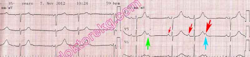
ECG 2. 2:1 AV block in a patient with coronary artery disease. Blocked P waves are shown by arrows.
Narrow QRS complexes suggest Wenkebach type 2:1 AV block.
Click here for a more detailed ECG

ECG 3. Holter recording shows Wenkebach phenomenon. PR interval gradually prolongs and
the 5th beat from the left is not conducted . The cycle restarts and PR interval gradually prolongs again .
Dr. Gulay Copur has donated the above ECG to our website.
Click here for a more detailed ECG
ECG 4. The ECG above belongs to a 65 years-old woman complaining of dizziness. There is Mobitz Type 1 (Wenkebach) AV
block. After a non-conducted (dropped) P wave the cycle restarts and PR interval prolongs gradually .
The third P wave fails to be conducted again (dropped beat) .
Dr. Nezire Gullu has donated the above ECG to our website.
Click here for a more detailed ECG

ECG 5. The ECG above belongs to a 6 months-old baby with a large, non-restrictive VSD.
Congenital 2:1 atrioventricular block with ventriculophasic sinus arrhythmia is seen.
Pediatric cardiologist Dr. Mahmut Gokdemir has donated the above ECG to our website.
Click here for a more detailed ECG
Diagnostic criteria
PR interval is not fixed. There is gradual prolongation of the PR interval, and after a few
beats one of the P waves cannot be conducted to the ventricles. Thereafter, the cycle restarts.
Some P waves are not followed by a QRS complex (
dropped beat).
Block is at the level of atrioventricular (AV) node.
2:1 AV block is generally accepted as a variant of the Wenkebach phenomenon.
ECG 1. Gradual prolongation of the PR interval. Blocked (non-conducted) P waves are shown by arrows.
Click here for a more detailed ECG
ECG 2. 2:1 AV block in a patient with coronary artery disease. Blocked P waves are shown by arrows.
Narrow QRS complexes suggest Wenkebach type 2:1 AV block.
Click here for a more detailed ECG
ECG 3. Holter recording shows Wenkebach phenomenon. PR interval gradually prolongs and
the 5th beat from the left is not conducted . The cycle restarts and PR interval gradually prolongs again .
Dr. Gulay Copur has donated the above ECG to our website.
Click here for a more detailed ECG
ECG 4. The ECG above belongs to a 65 years-old woman complaining of dizziness. There is Mobitz Type 1 (Wenkebach) AV
block. After a non-conducted (dropped) P wave the cycle restarts and PR interval prolongs gradually .
The third P wave fails to be conducted again (dropped beat) .
Dr. Nezire Gullu has donated the above ECG to our website.
Click here for a more detailed ECG
ECG 5. The ECG above belongs to a 6 months-old baby with a large, non-restrictive VSD.
Congenital 2:1 atrioventricular block with ventriculophasic sinus arrhythmia is seen.
Pediatric cardiologist Dr. Mahmut Gokdemir has donated the above ECG to our website.
Click here for a more detailed ECG